A.N. Belousov1,2*, E.Yu. Belousova1, A.V. Mysyk3
1Laboratory of Applied Nanotechnology of Belousov
2Kharkiv National Medical University, Ukraine
3Krasnokutsk Central District Hospital, Ukraine
*Correspondence: A.N. Belousov, Laboratory of Applied Nanotechnology of Belousov, 2Kharkiv National Medical University, Ukraine. E-mail: an.belousov2012@ukr.net
Copyright: © 2024 A.N. Belousov. This is an open-access article distributed under the terms of the Creative Commons Attribution License, which permits unrestricted use, distribution, and reproduction in any medium, provided the original author and source are credited.
Citation: Belousov, A. N, Belousova E. Yu, and Mysyk A.V. “Therapeutic Use of Biocompatible Nanodevices In Multiple Sclerosis: Results of A Clinical Trial.” J Nanotech Nanobiotech (2025): 105. DOI: 10.59462/JNNB.1.1.101.
Received date: 21 November, 2024; Accepted date: 23 December, 2024; Published date: 30 December, 2024
Multiple sclerosis (MS) is a significant neurological disorder due to its high prevalence, chronic progression, frequent development of disability, and predominant onset in young individuals. The pathogenesis of MS is primarily explained by the immunopathogenesis hypothesis. Biocompatible magnetite nanoparticles, known for their selective sorption of cell membrane surface proteins, circulating immune complexes, lymphocytotoxic antibodies, and complement system components, have demonstrated potential for immunocorrection. Additionally, these nanoparticles enhance phagocytic activity and improve the leukocyte phagocytosis index, making them promising candidates for MS therapy. The primary objective of this study is to slow MS progression, improve neurological function and overall patient well-being, and mitigate the expansion of demyelinating lesions in the brain. Materials and Methods: The study included a patient diagnosed with secondary progressive multiple sclerosis in its cerebrospinal form during a phase of clinical exacerbation. Neurological status and disability levels were assessed using the Expanded Disability Status Scale (EDSS), and contrast-enhanced brain MRI was performed. The nanodevice Micromage-B was administered orally as an immunosorbent and immunomodulator, with dosage and treatment regimens tailored to the patient. Assessments of general condition and neurological function were conducted weekly over a six-month period, with a contrast-enhanced MRI scan performed in the fifth month. Results: Treatment with Micromage-B resulted in a measurable improvement in neurological status. Patient experienced reduced stiffness and fatigue in the lower limbs, improved gait and coordination, diminished hand tremors, alleviation of depression and concentration difficulties, restoration of appetite, and enhanced speech function. Throughout the treatment, positive trends in neurological normalization were observed. After six months, the total EDSS score decreased from 210 to 45. The most significant improvements were noted in pyramidal function and coordination, with a reduction in the EDSS Disability Scale score from 6.0 to 5.0. Remarkably, by the fourth month of treatment, contrast-enhanced MRI revealed a decrease in the number of newly formed demyelinating foci for the first time. The observed improvements in neurological status corresponded with MRI findings. The recovery of central nervous system function in MS appears to result not only from the immunosuppressive properties of magnetite nanoparticles but also from the activation of remyelination mechanisms and oligodendrocyte differentiation through enzymatic methylation. Conclusion: The use of biocompatible nanodevices in the complex treatment of MS is promising. Further improvement and study of the regimen and method of using biocompatible magnetite nanoparticles to enhance MS treatment effectiveness are required.
Keywords: Multiple sclerosis; Treatment; Nanodevice; Micromagе-B; Neurological status assessment; Remyelination.
Multiple sclerosis (MS) is a significant neurological issue due to its widespread prevalence, chronic nature, frequent progression to disability, and tendency to affect young people, with an average onset age of 30 years. The primary hypothesis of MS immunopathogenesis suggests that immunological tolerance is disrupted, leading to the active penetration of autoreactive cells sensitized to nervous tissue antigens through the blood-brain barrier into brain tissue. B-lymphocytes recognize myelin and signal T cells to initiate an immune attack [1-7]. T- and B-cells secrete chemicals that attract other immune cells, causing inflammation [8,9]. Plasma cells produce antibodies that attack myelin and recruit other immune cells [10,11]. T- and B-cells establish a persistent presence in the central nervous system (CNS) and continue their attack [12,13].
There are two main hypotheses for MS pathogenesis: the outside-in hypothesis, which posits that immunocompetent cells activated in the periphery penetrate brain tissue, and the inside-out hypothesis, which suggests primary damage to nervous tissue leads to the expression of damage receptors from the DAMPs (Danger-associated Molecular Patterns) family, resulting in immune activation and loss of tolerance to myelin antigens [14]. Antibodies produced by plasma cells actively destroy the protective covering of nerve cells, causing inflammation. Over time, this tissue becomes scarred, disrupting conduction. Consequently, impulses from the brain fail to reach the limbs and organs, leading to a loss of body control [15]. Common initial clinical symptoms include weakness and impaired sensation in one or more limbs, decreased vision, urinary disturbances, and cerebellar ataxia.
Currently, immunomodulatory and immunosuppressive drugs that alter the course of MS form the cornerstone of pathogenetic treatment. Their mechanisms of action involve: 1) selective immunosuppression, 2) complete immunosuppression, 3) prevention of the migration of activated cells from lymph nodes or into brain tissue, or 4) a combination of immunoregulatory, anti-inflammatory, antioxidant, and potentially neurotrophic actions. Future directions for new MS treatments include selective local immunocorrection, remyelination and neuroprotection, enhancement of neuroplasticity and functional relocalization, evaluation of the efficacy and safety of cell therapy, and individualized therapy selection based on predictions of pathological process variants and potential treatment responses derived from the molecular and cellular biology of MS [16-22].
The development of inflammation and demyelination in MS is driven by an impaired immune response, imbalance between regulatory and effector T cells, activation of B-cell immunity, and microglia. All drugs that impact the course of MS either deplete T- or B-cells or modify the signaling pathways involved in immune response formation [23].
Recently, significant attention has been given to the role of B-cells in chronic CNS inflammation, evidenced by their involvement in autoantibody formation, antigen presentation, and continuous T-cell activation in the brain parenchyma. Anti-B-cell therapies (Rituximab, Ocrelizumab, Ofatumumab) have shown effectiveness in both remitting and progressive MS. Stimulating remyelination in MS is associated with the development of new monoclonal antibody preparations, such as anti-LINGO and human immunoglobulin M (IgM), which promote remyelination and the differentiation of oligodendrocytes and their precursors [24].
One of the key mechanisms of axonal degeneration is mitochondrial dysfunction. Some drugs, such as Dimethylfumarate, Idebenone, and Biotin, show promise in addressing this issue [25]. Additionally, the use of drugs that affect the redistribution of ion channels in demyelinated axons (Lamotrigine, Amiloride, Fampridine) can help reduce the energy deficit in axons and neurons [26].
Recently, biocompatible nanotechnological preparations have seen increasing use in medicine. Since 1998, Ukrainian clinics have been officially using nanodevices developed by Belousov's Applied Nanotechnology Laboratory, including the Micromage-B, MCS-B, and ICNB brands (Figure 1) [27]. These nanodevices are based on biocompatible magnetite nanoparticles, whose unique physical and chemical characteristics enable a wide range of applications. They influence the quantitative and qualitative composition of bodily fluids, metabolic and biochemical processes, and the energy balance of cells. Their selective sorption activity against surface proteins of cell membranes, circulating immune complexes, lymphocytotoxic antibodies, and the complement system, as well as their ability to enhance phagocytic activity and leukocyte phagocytosis completion index [28], allow these nanodevices to be effectively used for immunocorrection. Moreover, these nanopreparations impact glycolysis processes, cell membrane ion channel activity, normalize erythrocyte function, improve microcirculation, and reduce the platelet aggregation index (Figure 2) [29-31]. They also activate the system of antiradical enzymes and inhibit lipid peroxidation processes [32,33].
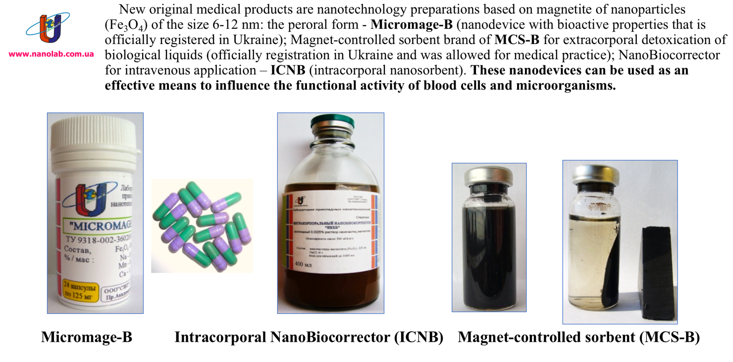 Figure 1. New official nanomedical devices based on biocompatible magnetite nanoparticles.
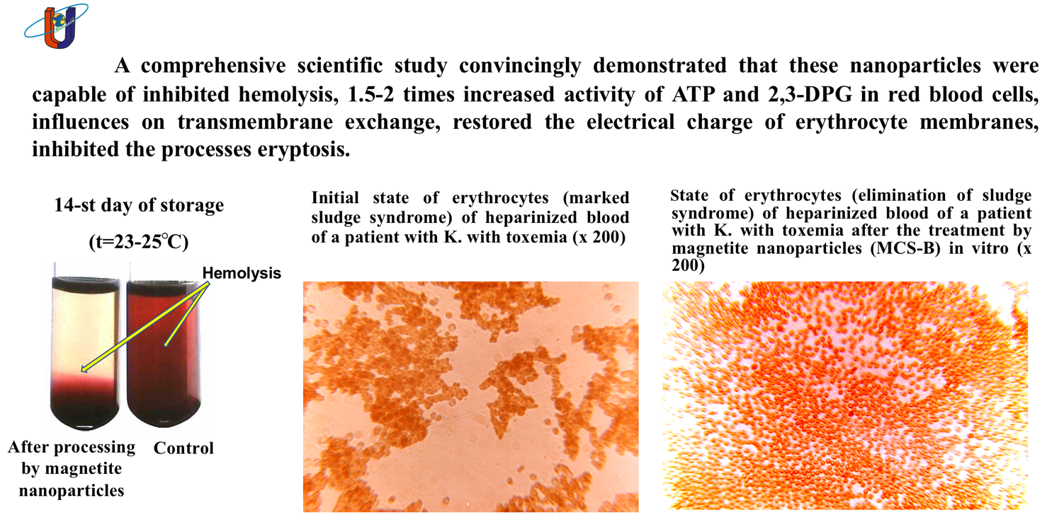
Figure 2. Effect of biocompatible magnetite nanoparticles on functional status of erythrocytes and hemorheology.
The results of these studies lay the foundation for developing innovative techniques for the effective and safe use of biocompatible magnetite nanoparticles in treating severe autoimmune diseases, including multiple sclerosis. The primary objective of the study is to slow the progression of MS, improve the neurological status and overall condition of the patient, and reduce the spread of demyelinating foci in the brain.
Patient K. was diagnosed with multiple sclerosis, secondary progressive type, cerebrospinal form, at the clinical aggravation stage. The patient presented with pronounced spastic tetraparesis, more severe in the lower extremities, leading to walking impairment, pronounced urinary-ataxic syndrome, and sphincter and sensory disorders. MRI revealed signs of multifocal diffuse brain lesions (more than 30) of a demyelinating nature, indicating the active phase of the disease, and a diffuse atrophic process in the cerebral cortex. The average number of relapses in the year before study inclusion was 1.0, and the EDSS disability score was 6.0. The disease had progressed for 24 years since the onset of the first symptoms. For 14 years, the patient received regular treatment with vascular and metabolic drugs in combination with hormonal therapy. In the 6th year, despite active treatment, the disease progressed to a secondary progressive course, leading to the addition of the immunosuppressive drug Teriflunomide to the therapy regimen.
However, despite ongoing treatment efforts, the patient’s general condition continued to deteriorate, and the neurological status remained unstable. MRI scans over the past four years showed a progressive increase in the number of new demyelinating lesions in the brain.
Given the above, the therapy regimen was supplemented with the prescription of Micromage-B [34]. Before starting the study, the patient's formal consent to the use of Micromag-B was obtained. This fact was recorded in the medical history, and the plan of treatment and subsequent study was agreed with the attending physician. Micromage-B is an oral nanodevice officially registered by the Ministry of Health of Ukraine. It is a powder form of magnetite (Fe3O4) nanoparticles designed for the prevention and treatment of various diseases and to enhance the body's resistance to adverse environmental factors. As a nanotechnology device, Micromage-B features magnetite nanoparticles sized between 6 and 12 nm. The therapeutic effect of Micromage-B is based on the mechanism of sorption and the action of a constant magnetic field on cellular and subcellular structures induced by the magnetite nanoparticles. The nanoparticles have a sorption surface area of 800 to 1200 m²/g and an induced magnetic field strength of 300-400 kA/m. The target of Micromage-B is the microenvironment of the cell's aqueous spaces and surface membrane proteins. Through selective sorption, the magnetite nanoparticles alter the quantitative and qualitative composition of cell surface proteins, and the constant magnetic field changes the mobility and orientation of hydrogen protons in the cell's aqueous microenvironment (Figure 3).
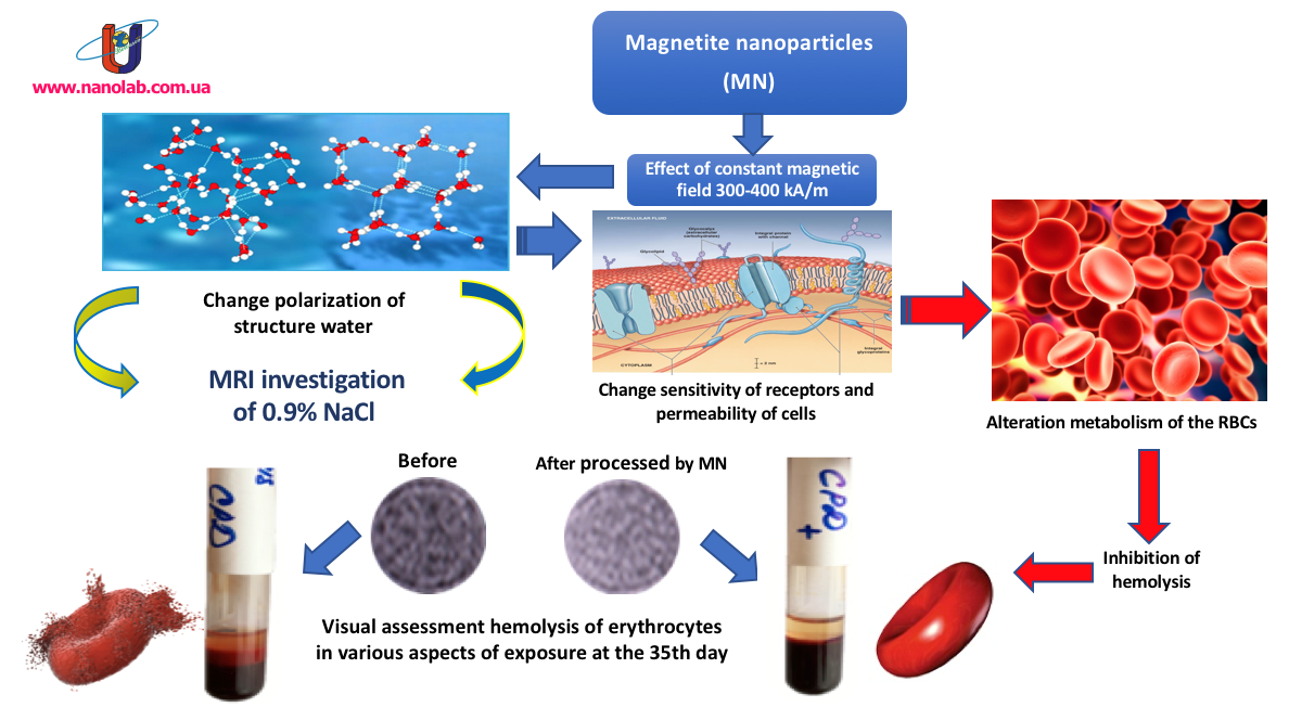
Figure 3. The effect of magnetite nanoparticles on hemolysis processes by changing the polarization structure of the aqueous sector of the erythrocyte microenvironment.
This leads to the activation of hydrolysis of the phosphate residue ATP, ultimately modifying the cell's transmembrane exchange and metabolism, and altering its susceptibility. The nanodevice enhances adaptive mechanisms and the potential of cell organelles, accelerates reparative processes at the membrane and macromolecule levels, significantly increases the level of synthetic intracellular reactions. Additionally, Micromage-B nanoparticles adsorb toxic substances and circulating immune complexes, significantly enhancing the treatment efficacy of various allergic diseases, autoimmune processes (such as rheumatoid arthritis, acute and chronic polyarthritis, eczema, etc.), and acute poisoning. Micromage-B regulates the activity of antioxidant enzymes, absorbs lipid peroxidation products, and rebalances antiradical and pro-radical product levels [35,36].
The dosing regimen for Micromage-B entails 500 mg daily for the first month, 500 mg every other day for the second month, and subsequently, 500 mg once every three days. The selection of Micromage-B dosage and regimen is tailored individually, considering the patient's rate of improvement and neurological recovery.
The study monitored changes in neurological status using a modified version of the Multiple Sclerosis Patient Assessment Scale [37,38], which evaluates the severity of motor impairments alongside other nervous system damage indicators. Disability was quantitatively assessed using Kurtzke's online EDSS calculator [39]. Manifestations of cerebral demyelination foci were examined through contrast-enhanced MRI.
The patient's overall condition and neurological status were assessed every 7 days over a 6-month period. As per the protocol, a contrast-enhanced MRI of the brain was conducted annually, aligning with the 5th month of Micromage-B usage.
The progression of neurological changes was assessed using a modified scale. One week following the administration of Micromage-B, a marked enhancement in the patient's neurological condition was evident. The patient reported a notable reduction in lower limb stiffness and rapid fatigue. Objectively, improvements were observed in gait and coordination, reduction in hand tremors, complete alleviation of depression and concentration issues, restoration of appetite, and enhancement in speech. Positive trends in normalizing neurological status persisted throughout the entire duration of Micromage-B treatment. Table 1 illustrates the evaluation of a multiple sclerosis patient's neurological status before and after 6 months of Micromage-B administration.
|
1. Movements (pyramidal system) |
|||||
|
Input data |
Six months after application Micromage-B |
Clinical manifestations |
|||
|
Arm |
Leg |
Arm |
Leg |
||
|
0 |
0 |
0 |
0 |
Norma |
|
|
5 |
10 |
5* |
10* |
Absence of loss symptoms, revival of tendon reflexes, enlargement of reflexogenic zones, clonus, presence of pathological signs, anisoreflexia, (absence of paresis) |
|
|
10 |
20 |
10 |
20 |
Raises a limb independently, full volume of active movements, signs of pyramidal lesion, overcomes not only the gravity of his own limb, but also an additional obstacle of moderate strength, positive Barre-Rusetsky's symptom |
|
|
15 |
40 |
15 |
40 |
Raises a limb independently, the volume of active movements is full, cannot hold a limb in a given position for a long time, and cannot overcome an additional obstacle. |
|
|
20* |
60* |
20 |
60 |
Can pull a limb off the plane, the amount of active movement is limited, cannot hold a limb in a given position. |
|
|
40 |
80 |
40 |
80 |
Cannot detach a limb from the plane, active movements in the joints of the fingers, ankle and wrist, elbow, knee joints are possible only on the plane. |
|
|
50 |
100 |
50 |
100 |
Complete absence of movement (paralysis) |
|
|
2. Sensitivity |
|||||
|
Before |
After |
(a) superficial sensitivity |
|||
|
0 |
0* |
Norma |
|||
|
5 |
0 |
Paresthesias, burning sensation, numbness, coldness of a limb (no objective disorders) |
|||
|
10* |
10 |
Hyperesthesia or hypoesthesia |
|||
|
15 |
15 |
Anesthesia phenomena |
|||
|
|
b) deep sensitivity |
||||
|
0* |
0* |
Norma |
|||
|
10 |
10 |
Disorder of joint and muscle feeling in small joints |
|||
|
20 |
20 |
Disorder of joint and muscle feeling up to the level of the middle joints (wrist, ankle) |
|||
|
40 |
40 |
Disorder of joint and muscle feeling up to the level of large joints (elbow, shoulder, knee, hip) |
|||
|
3. Coordination |
|||||
|
0 |
0 |
Norma |
|||
|
10 |
10* |
Unsteadiness, swaying in the sensitized Romberg's test while standing on one leg, mild intensional trembling (in the complicated test), slight ataxia in the heel-knee test, deviation when walking with eyes closed. |
|||
|
40* |
10 |
Unsteadiness in simple Romberg's pose, atactic gait with open eyes and legs spread wide apart, "drunkenness," moderately pronounced intensional tremor and ataxia in the heel-knee test. |
|||
|
100 |
100 |
Because of ataxia, the patient cannot move without assistance, sharp hypotonia of muscles, intensional shaking of the head, trunk, coarse - upper extremities, coarse ataxia during heel-knee test, trembling of upper extremities when trying to perform purposeful movement, chanted speech. |
|||
|
4. Psycho-emotional sphere |
|||||
|
0 |
0* |
Norma |
|||
|
10 |
10 |
Mild impairment of the intellect in combination with euphoria, rapid change of mood, neurasthenic syndrome. |
|||
|
20* |
0 |
Euphoria, depression, decreased criticism of one's condition, decreased memory. |
|||
|
100 |
100 |
Severe mental disorder, complete intellectual disintegration, Korsak's syndrome, etc. |
|||
|
5. Nystagmus |
|||||
|
0 |
0* |
Norma |
|||
|
5 |
5 |
Nystagmus is detected only in the extreme leads (degree 1) |
|||
|
10* |
10 |
Nystagmus when looking straight ahead (degree 2) |
|||
|
15 |
15 |
Sharp beating nystagmus, nystagmus in both directions, even toward the slow component (3rd degree) |
|||
|
|||||
|
0 |
0* |
Norma |
|||
|
10* |
10 |
Impulsive urges, inability to hold urine for a long time, difficulty urinating |
|||
|
20 |
20 |
Urinary incontinence, urinary retention, intermittent urination disorders, persistent constipation |
|||
|
7. Sexual function |
|||||
|
0 |
0* |
Norma |
|||
|
5* |
5 |
Decreased sexual activity in men (intermittent impotence), sexual coldness in women |
|||
|
10 |
10 |
Total impotence, menstrual disorders in women |
|||
|
The ocular fundus |
|||||
|
0 |
0 |
Norma |
|||
|
5 |
5 |
Disturbance of vascular pattern, narrowing of arteries, dilation of veins, changes on fluorescence ophthalmoscopy |
|||
|
10* |
10* |
Partial optic atrophy (bitemporal pallor), optic neuritis |
|||
|
15 |
15 |
Complete atrophy of the optic nerve |
|||
|
|||||
|
0 |
0 |
Norma (vision within 1.0 or myopia) |
|||
|
5 |
5 |
Occasional blurring of vision, rapid fatigue when reading and performing work without impaired visual acuity |
|||
|
10* |
10* |
Visual acuity from 0.9 to 0.7 |
|||
|
15 |
15 |
Visual acuity from 0.6 to 0.4 |
|||
|
20 |
20 |
Visual acuity from 0.3 to 0.1 |
|||
|
25 |
25 |
Visual acuity 0.1 and below |
|||
|
100 |
100 |
Blindness in one or both eyes |
|||
|
|||||
|
0 |
0* |
Norma (absence of subjective and objective symptoms) |
|||
|
5* |
0 |
Concealed insufficiency, without visible dysfunction of one of the oculomotor nerves, inter-nuclear ophthalmoplegia syndrome |
|||
|
10 |
10 |
Mild visible impairment, visible insufficiency of one or more nerves, diplopia, ptosis, anisocoria |
|||
|
15 |
15 |
Convergent or divergent strabismus |
|||
|
20 |
20 |
Complete ophthalmoplegia (in one or both eyes) |
|||
|
11. The trigeminal nerve |
|||||
|
0* |
0* |
Norma (absence of subjective and objective symptoms) |
|||
|
5 |
5 |
Subjective sensations in the form of pain, numbness, sense of "creeping chills", oppression in the face. |
|||
|
10 |
10 |
Objective signs of lesions, hypoesthesia, loss or decrease in the corneal reflex. |
|||
|
20 |
20 |
Severe anomalies with loss of trigeminal nervous functions, with or without neuralgic disorders. |
|||
|
12. The facial nerve |
|||||
|
0 |
0* |
Norma |
|||
|
5* |
0 |
Moderate weakness of facial muscles (eye closes completely, but cannot actively close it), asymmetry of frontal and nasolabial folds |
|||
|
10 |
10 |
Moderate weakness of mimic muscles (lagophthalmus, positive Bell's symptom, facial asymmetry in grinning), with preservation, to some extent, of mimic movements |
|||
|
20 |
20 |
Complete paralysis of facial muscles |
|||
|
|||||
|
0 |
0* |
Norma |
|||
|
5* |
5 |
Mildly pronounced bulbar phenomena (gagging when taking liquid food, change in speech, without gross organic symptoms of prolapse) |
|||
|
10 |
10 |
Severe dysphagia, dysarthria, decreased soft palate and posterior pharyngeal wall reflexes |
|||
|
100 |
100 |
Complete bulbar paralysis |
|||
|
|||||
|
0* |
0* |
Norma |
|||
|
5 |
5 |
Conversational speech at a distance of 4 to 6 m, whispered speech at a distance of 1 to 3 m. |
|||
|
10 |
10 |
Speech - from 2 to 4 m, whisper - from 0.5 to 1 m. |
|||
|
15 |
15 |
Spoken speech 2 m or less, whispered speech 0 to 0.5 m |
|||
|
20 |
20 |
Complete deafness in one or both ears |
|||
|
Total: |
Total: |
|
|||
|
210* |
45* |
|
|||
Table 1. Assessment of Neurological Status Scale for Patient K. with Multiple Sclerosis.
Note*: estimated scores of the neurological status before and after using the Micromage-B.
The data presented in Table 1 illustrate a positive trend in normalizing neurological status following 6 months of Micromage-B usage. Initially, the total points amounted to 210, which decreased to 45 after the 6-month period, indicating a reduction of 165 points. The most significant improvement was observed in the assessment of the pyramidal system and coordination. Additionally, the EDSS disability scale score decreased from 6.0 to 5.0.
A contrast-enhanced MRI of the brain conducted after 4 months of Micromage-B usage revealed a decrease in the number of new demyelination foci in the brain for the first time. The favorable progression of neurological status correlated with the brain MRI findings.
Upon analyzing the collected data, particular attention should be given to the observed positive clinical effects attributed to immunosorption and the active contribution of Micromage-B's magnetite nanoparticles to neurological status restoration. This effect may be attributed to remyelination processes and oligodendrocyte differentiation. Oligodendrocytes, a type of neuroglial cell, form the myelin sheath around neurons in the central nervous system (CNS). The molecular mechanisms underlying cell differentiation and specialization remain complex and poorly understood, presenting a challenging area in cell and developmental biology. The development and maturation of various cell types continue to pose significant research challenges.
However, it is now understood that one mechanism underlying oligodendrocyte maturation involves enzymatic methylation, specifically the addition of a methyl group (-CH3) to the N6 nitrogen atom in adenosine's nitrogenous base, referred to as m6A-methylation. Despite its seemingly minor nature, this modification can significantly impact subsequent stages of protein biosynthesis. The role of m6A-methylation has been demonstrated in numerous processes associated with oligodendrocyte maturation [40]. The body's universal methyl group donor is S-adenosylmethionine, generated through the interaction between the amino acid methionine and ATP.
Given that Micromag-B activates glycolysis, it significantly increases the production of the macroergic compound ATP [41,42], and promotes the formation of the reduced form of the coenzyme NADPH2, facilitating the conversion of oxidized glutathione to its reduced form [43]. These conditions are conducive to initiating enzymatic methylation processes, which likely enhance the mechanism of action of magnetite nanoparticles (Micromag-B). This, in turn, supports the differentiation of oligodendrocytes and the remyelination process. Additionally, it should be noted that these nanoparticles polarize the aqueous environment of the cellular microenvironment [44], leading to the activation of ATP hydrolysis, energy release, and the formation of ADP. For a detailed understanding of how biocompatible magnetite nanoparticles (Micromag-B) influence remyelination processes, refer to (Figure 4).
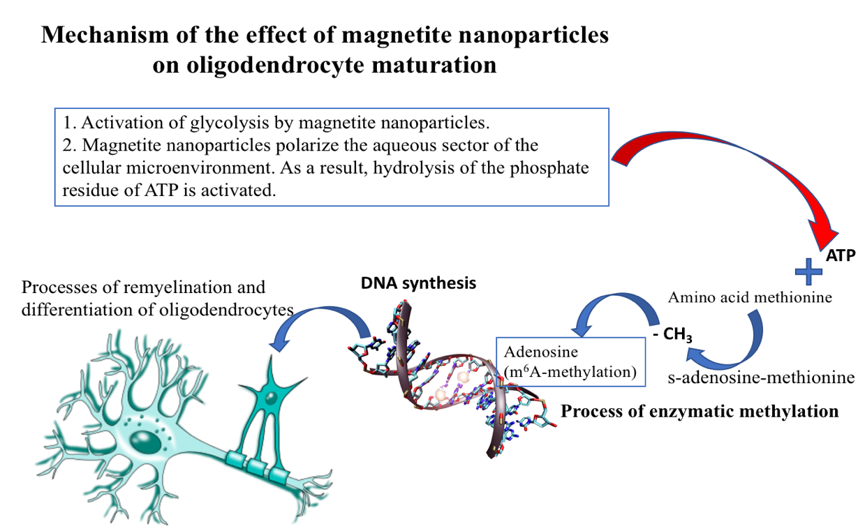
Figure 4. Mechanism of the effect of magnetite nanoparticles on oligodendrocyte maturation.
Figure 4. Mechanism of the effect of magnetite nanoparticles on oligodendrocyte maturation.
Given the positive neurological status dynamics, it was deemed appropriate to continue Micromage-B nanopreparation administration at the prescribed dosage. Therapy was augmented with a regimen of rehabilitative exercises aimed at expediting the restoration of physical, cognitive, and psychosocial functions in patients with MS.
Considering the positive progression in neurological status, it was decided to continue the administration of the nanopreparation Micromage-B at the prescribed dosage. Additionally, the therapy was augmented with a comprehensive rehabilitation exercise program designed to expedite the recovery of physical, cognitive, and psychosocial functions in a patient with MS.
The study's results broadened the clinical effectiveness of biocompatible magnetic nanoparticles in treating severe autoimmune diseases [45-48]. The use of the Micromage-B nanopreparation in treating multiple sclerosis (MS) demonstrated a significant positive clinical effect. Positive trends in normalizing the neurological state were observed throughout the application period of Micromage-B. After six months of treatment, the overall score decreased from 210 to 45. The most notable improvement was seen in the assessment of the pyramidal system and coordination. The EDSS Disability Scale score decreased from 6.0 to 5.0. For the first time, contrast-enhanced MRI of the brain recorded a reduction in the number of new demyelination foci by the fourth month of Micromage-B administration. The normalized neurological condition correlated with positive brain MRI results. The restoration of central nervous system activity in MS is attributed not only to the immunosuppressive properties of magnetite nanoparticles but also likely to the activation of remyelination mechanisms and oligodendrocyte differentiation through enzymatic methylation. The design and method of using biocompatible magnetite nanoparticles to enhance MS treatment efficiency require further refinement and research. The use of biocompatible nanodevices in the comprehensive treatment of MS is a promising innovation [49-51]. Micromage-B is a biologically active form of a nanodevice officially approved by the Ministry of Health of Ukraine and authorized for clinical use since 1998. To date, no side effects or contraindications to its use have been identified. Given the key properties of biocompatible magnetite nanoparticles, their clinical applications continue to expand. Recent studies have deepened our understanding of the mechanisms regulating cellular metabolism, microcirculation, eryptosis, as well as the mechanisms of action of antibacterial agents and other processes. To substantiate the necessity of further utilizing magnetite nanoparticles in larger clinical trials, it is crucial first to identify relevant biochemical markers. These markers should not only objectively reflect the severity of multiple sclerosis from a pathogenetic perspective but also provide a high-probability prognosis of disease outcomes.
Duffy, Samuel S., Justin G. Lees, and Gila Moalem-Taylor. "The contribution of immune and glial cell types in experimental autoimmune encephalomyelitis and multiple sclerosis." Multiple sclerosis international 2014, no. 1 (2014): 285245.
Ortiz, Genaro Gabriel, Fermín Paul Pacheco-Moisés, Miguel Ángel Macías-Islas, and Luis Javier Flores-Alvarado, et al. "Role of the blood–brain barrier in multiple sclerosis." Archives of medical research 45, no. 8 (2014): 687-697.
Larochelle, Catherine, Jorge Ivan Alvarez, and Alexandre Prat. "How do immune cells overcome the blood–brain barrier in multiple sclerosis?." FEBS letters 585, no. 23 (2011): 3770-3780.
Cross, Anne H., and Emmanuelle Waubant. "MS and the B cell controversy." Biochimica et Biophysica Acta (BBA)-Molecular Basis of Disease 1812, no. 2 (2011): 231-238.
Dalakas, Marinos C. "B cells as therapeutic targets in autoimmune neurological disorders." Nature clinical practice Neurology 4, no. 10 (2008): 557-567.
Constant, Stephanie L. "B lymphocytes as antigen-presenting cells for CD4+ T cell priming in vivo." The Journal of Immunology 162, no. 10 (1999): 5695-5703.
Crawford, Alison, Megan MacLeod, Ton Schumacher, Louise Corlett, and David Gray. "Primary T cell expansion and differentiation in vivo requires antigen presentation by B cells." The Journal of Immunology 176, no. 6 (2006): 3498-3506.
Bar‐Or, Amit, Lama Fawaz, Boli Fan, Peter J. Darlington, Aja Rieger, Christine Ghorayeb, Peter A. Calabresi et al. "Abnormal B‐cell cytokine responses a trigger of T‐cell–mediated disease in MS?." Annals of neurology 67, no. 4 (2010): 452-461.
Duddy, Martin, Masaaki Niino, Femina Adatia, Sherry Hebert, Mark Freedman, Harry Atkins, Ho Jin Kim, and Amit Bar-Or. "Distinct effector cytokine profiles of memory and naive human B cell subsets and implication in multiple sclerosis." The Journal of Immunology 178, no. 10 (2007): 6092-6099.
CP, GENAIN. "Identification of autoantibodies associated with myelin damage in multiple sclerosis." Nat. Med. 5 (1999): 170.
Storch, Maria K., Sara Piddlesden, Matti Haltia, Matti Iivanainen, Paul Morgan, and Hans Lassmann. "Multiple sclerosis: in situ evidence for antibody‐and complement‐mediated demyelination." Annals of neurology 43, no. 4 (1998): 465-471.
Serafini, Barbara, Barbara Rosicarelli, Roberta Magliozzi, Egidio Stigliano, and Francesca Aloisi. "Detection of ectopic B‐cell follicles with germinal centers in the meninges of patients with secondary progressive multiple sclerosis." Brain pathology 14, no. 2 (2004): 164-174.
Magliozzi, Roberta, Owain W. Howell, Cheryl Reeves, Federico Roncaroli, Richard Nicholas, Barbara Serafini, Francesca Aloisi, and Richard Reynolds. "A gradient of neuronal loss and meningeal inflammation in multiple sclerosis." Annals of neurology 68, no. 4 (2010): 477-493.
Dhaiban, Sarah, Mena Al-Ani, Noha Mousaad Elemam, Mahmood H. Al-Aawad, Zeinab Al-Rawi, and Azzam A. Maghazachi. "Role of peripheral immune cells in multiple sclerosis and experimental autoimmune encephalomyelitis." Sci 3, no. 1 (2021): 12.
Kobelt, Gisela, Alan Thompson, Jenny Berg, Mia Gannedahl, Jennifer Eriksson, MSCOI Study Group, and European Multiple Sclerosis Platform. "New insights into the burden and costs of multiple sclerosis in Europe." Multiple Sclerosis Journal 23, no. 8 (2017): 1123-1136.
Farooqi, Nasr, Bruno Gran, and Cris S. Constantinescu. "Are current disease‐modifying therapeutics in multiple sclerosis justified on the basis of studies in experimental autoimmune encephalomyelitis?." Journal of neurochemistry 115, no. 4 (2010): 829-844.
Polman, Chris H., Paul W. O'Connor, Eva Havrdova, Michael Hutchinson, Ludwig Kappos, David H. Miller, J. Theodore Phillips et al. "A randomized, placebo-controlled trial of natalizumab for relapsing multiple sclerosis." New England Journal of Medicine 354, no. 9 (2006): 899-910.
Yednock, Ted A., Catherine Cannon, Lawrence C. Fritz, Francisco Sanchez-Madrid, Lawrence Steinman, and Nathan Karin. "Prevention of experimental autoimmune encephalomyelitis by antibodies against α 4 β l integrin." Nature 356, no. 6364 (1992): 63-66.
Ridge, Susan C., Adolph E. Sloboda, Richard A. McReynolds, Seymour Levine, Arnold L. Oronsky, and S. S. Kerwar. "Suppression of experimental allergic encephalomyelitis by mitoxantrone." Clinical immunology and immunopathology 35, no. 1 (1985): 35-42.
Huang, Wen-Juan, Wei-Wei Chen, and Xia Zhang. "Multiple sclerosis: Pathology, diagnosis and treatments." Experimental and therapeutic medicine 13, no. 6 (2017): 3163-3166.
Ghasemi, Nazem, Shahnaz Razavi, and Elham Nikzad. "Multiple sclerosis: pathogenesis, symptoms, diagnoses and cell-based therapy." Cell Journal (Yakhteh) 19, no. 1 (2016): 1.
Hanafy, Khalid A., and Jacob A. Sloane. "Regulation of remyelination in multiple sclerosis." FEBS letters 585, no. 23 (2011): 3821-3828.
Ghasemi, Nazem, Shahnaz Razavi, and Elham Nikzad. "Multiple sclerosis: pathogenesis, symptoms, diagnoses and cell-based therapy." Cell Journal (Yakhteh) 19, no. 1 (2016): 1.
Høglund, Rune A., and Azzam A. Maghazachi. "Multiple sclerosis and the role of immune cells." World journal of experimental medicine 4, no. 3 (2014): 27.
Barcelos, Isabella Peixoto de, Regina M. Troxell, and Jennifer S. Graves. "Mitochondrial dysfunction and multiple sclerosis." Biology 8, no. 2 (2019): 37.
Hamada, Mustafa S., and Maarten HP Kole. "Myelin loss and axonal ion channel adaptations associated with gray matter neuronal hyperexcitability." Journal of Neuroscience 35, no. 18 (2015): 7272-7286.
A.N. Belousov. Nanotechnology is a key priority for the foreseeable future in medicine.
Belousov, Аndrey Nikolaevich. "The use of magnetite nanoparticles in applied medicine." In Materials Science Forum, vol. 694, pp. 205-208. Trans Tech Publications Ltd, 2011.
Belousov, Andrey. "The role of magnetite nanoparticles (ICNB) in discovery new factor which influence on permeability of erythrocytes and eryptosis." Open Access Library Journal 1, no. 8 (2014): 1-6.
Belousov, A. N., Elena Malygon, Vadim Yavorskiy, and Ekateryna Belousova. "Application of the standardized form magnetite nanoparticles (ICNB) in creature simple and practical method of additive modernization of preservation solutions for red blood cells." Global Journal of Anesthesia and Pain Medicine 1, no. 1 (2018): 1.
Belousov, A. "Study of the Effect of Nanotechnology Drugs (mcs-b) on the Aggregation of Human Blood Platelets." Journal of Nanosciences Research & Reports. Scientize Publishers 1, no. 1 (2020): 1-8.
Belousov, Andrey, Elena Malygon, Vadim Yavorskiy, and Ekateryna Belousova. "Innovative method of nanotechnology to increase the storage time of RBCs due by stabilizing the molecular structure of proteins and lipids of erythrocyte membranes." Biomedical Journal 1 (2019): 10.
Belousov, A., T. Kalynychenko, E. Malygon, M. Anoshyna, M. Yagovdik, V. Yavorskiy, and E. Belousova. "Research of Lipid Peroxidation after Administration of Nanomodified Resuspending Solution in Donor Red Blood Cells on During Their Storage." International Journal of Biomed Research 2, no. 2 (2022).
Patent Agency of Ukraine No. 30538А UA A 23L 1/304 Therapeutic and preventive product MICROMAGE-B / A.N. Belousov № 98052704. 25.05.98. Publ. 15.11.00. Bul. No. 6-11.
Andrey N. Belousov. “Extracorporal hemocorrection using magnet-controlled sorbent in intensive therapy of intoxication syndromes in patients with hepatopancreatoduodenales diseases”.
Belousov, Andrey. "A New Promising Method of Hepatitis Treatment at the Level of Ultrastructure of the Liver by Standardized Powder form of Magnetite Nanoparticles (Micromage-B)." Редакційна колегія (2019): 8.
Gordeev, Y. Y., T. M. Shamova, and V. V. Semashko. "Scale of evaluation of the neurological status in multiple sclerosis.(2006)." Journal of GrSMU 1: 75-78.
Kurtzke, John F. "Rating neurologic impairment in multiple sclerosis: an expanded disability status scale (EDSS)." Neurology 33, no. 11 (1983): 1444-1444.
Xu, Huan, Yulia Dzhashiashvili, Ankeeta Shah, Rejani B. Kunjamma, Yi-lan Weng, Benayahu Elbaz, Qili Fei et al. "m6A mRNA methylation is essential for oligodendrocyte maturation and CNS myelination." Neuron 105, no. 2 (2020): 293-309.
Belousov, A. N. "Effect on hemolysis and transport ATPase activity of erythrocytes by means nanopareticles of magnetit controlled sorbent (MCS-B)." Pain, anesthesia and intensive care. Kiev 1 (2012): 26-8.
Belousov, Andrey. "The role of magnetite nanoparticles (ICNB) in discovery new factor which influence on permeability of erythrocytes and eryptosis." Open Access Library Journal 1, no. 8 (2014): 1-6.
Belousov, Andrey, Tetiana Kalynychenko, Elena Malygon, Militina Anoshyna, Maryna Yagovdik, Vadim Yavorskiy, and Ekateryna Belousova. "Study of effects a new resuspending solution which was nanotechnologically upgraded on lipoperoxidation, catalase activity and red blood cell peroxidation resistance in donor blood components." Archives in Biomedical Engineering & Biotechnology 6, no. 2 (2021): 1-6.
Belousov, A. N. "Myth and reality application of magnetite nanoparticles as selective contrasting means of the malignant tumors in MRI investigation." Journal Biomedical Engineering Research 2, no. 3 (2014): 147-152.
Singh, Ajay Vikram, Manish Khare, W. N. Gade, and Paolo Zamboni. "Theranostic implications of nanotechnology in multiple sclerosis: a future perspective." Autoimmune diseases 2012, no. 1 (2012): 160830.
Silva, Gabriel A. "Neuroscience nanotechnology: progress, opportunities and challenges." Nature reviews neuroscience 7, no. 1 (2006): 65-74.
Kanwar, Jagat R., Xueying Sun, Vasu Punj, Bhasker Sriramoju, Rajiv R. Mohan, Shu-Feng Zhou, Ashok Chauhan, and Rupinder K. Kanwar. "Nanoparticles in the treatment and diagnosis of neurological disorders: untamed dragon with fire power to heal." Nanomedicine: Nanotechnology, Biology and Medicine 8, no. 4 (2012): 399-414.
Srikanth, Maya, and John A. Kessler. "Nanotechnology—novel therapeutics for CNS disorders." Nature reviews neurology 8, no. 6 (2012): 307-318.
Fazil, Mohammad, Shadab, Sanjula Baboota, Jasjeet K. Sahni, and Javed Ali. "Nanotherapeutics for Alzheimer’s disease (AD): past, present and future." Journal of drug targeting 20, no. 2 (2012): 97-113.
Wankhede, Mamta, Alexandros Bouras, Milota Kaluzova, and Costas G. Hadjipanayis. "Magnetic nanoparticles: an emerging technology for malignant brain tumor imaging and therapy." Expert review of clinical pharmacology 5, no. 2 (2012): 173-186.
Patel, Toral, Jiangbing Zhou, Joseph M. Piepmeier, and W. Mark Saltzman. "Polymeric nanoparticles for drug delivery to the central nervous system." Advanced drug delivery reviews 64, no. 7 (2012): 701-705.