Eduardo D. Rubio1* and Mariano Mombru2
Chairman Oral and Maxillofacial Surgery Training Program, Graduate School of Medical Sciences, Pontificia Universidad Católica, Argentina.
Adjunct Professor, Oral and Maxillofacial Surgery Training Program, Graduate School of Medical Sciences, Pontificia Universidad Católica, Argentina.
Received: 01 April 2024; Accepted: 14 May 2024; Published: 21 May 2024
Citation: Rubio Eduardo D. and Mombru Mariano. “A New Classification System for Deeply Impacted Third Mandibular Molars.” J Oral Dis Treat (2024): 104. DOI: 10.59462/JODT.1.1.104
Copyright: © 2024 Rubio ED. This is an open-access article distributed under the terms of the Creative Commons Attribution License, which permits unrestricted use, distribution, and reproduction in any medium, provided the original author and source are credited.
Third molars extractions are one of the most common procedures for an oral and maxillofacial surgeon (OMFS). In some cases, third mandibular molars extractions could be difficult regarding possible inferior alveolar nerve (IAN) injury. Moreover, mandible weakening due to excessive bone removal during the procedure, may led to bone defects and high risk of mandibular fracture (MF). The aim of this study is to propose a new classification system to assess IAN injury and MF risks during or after DITMM extraction, as well as the need of bone grafting in relation to the amount of bone removed during to the surgery.
IAN injury • Periodontal disease • DITMM • Bone grafting • Cone beam computed tomography
Although third molars extraction is one of the most common procedures in the dental office, in some cases, could be difficult regarding possible inferior alveolar nerve (IAN) injury [1]. Furthermore, mandible weakening because of bone removal, may led to bone defects and high risk of mandibular fracture (MF) during or after the procedure. Additionally, damage to adjacent teeth has been described as a complication of third mandibular molars extractions, among others. Hence, OMFS must chose an appropriate surgical technique when deeply impacted third mandibular molars (DITMM) should be extracted, due to infection or odontogenic pathologic conditions associated to the impacted tooth. At the time, there is no classification system of DITMM considering risk of IAN injury and MF.
A randomized - retrospective descriptive study was designed to assess the position of 100 third mandibular molars with CBCT scans including only, DITMM for this study. 238 CBCT scans of third mandibular molars from patients that consulted to our private practice between 2019 and 2023 were considered, in a randomized selection. 138 were excluded because of the presence of third mandibular molars not considered DITMM, lack of signs of infection and absence of pathologic condition associated to the DITMM, and thus, with no indication for extraction. An impacted third mandibular was consider as DITMM when the distance between the tip of the root and the inferior border of the mandible was at least or less than 10 mm, regardless of the occlusal plane of the second molar.
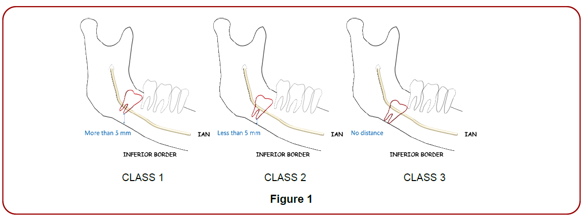
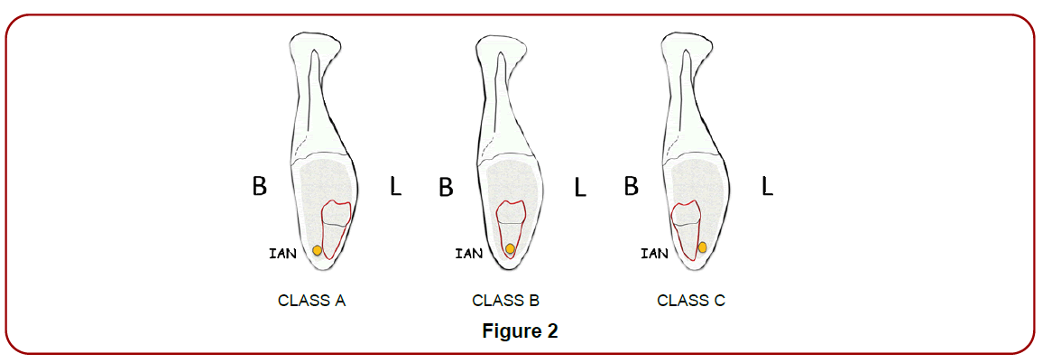
The presence or absence of signs of infection were assessed in the clinical record of each patient. The presence or absence of pathologic condition associated to the DITMM, was evident in each case, at the time of the CBCT scans evaluation. The distance between the tip of the root and the inferior border of the mandible was measured with Blue Sky Plan 4 (Blue Scan Bio Software), and each molar was classified as class 1, 2 and 3 as show in (Figure 1) and (Table 1). Additionally, the position of IAN was assessed in CBCT frontal scans according to the location of the DITMM and classified as class A, B and C as show in (Figure 2) and (Table 2).

Figure 1.

Figure 2.
| Class 1 | The distance between the inferior border of the mandible and the tip of the dental roots of the third molar is more than 5 mm. |
| Class 2 | The distance between the inferior border of the mandible and the tip of the dental roots of the third molar is 5 mm or less. |
| Class 3 | There is no distance between the inferior border of the mandible and the tip of the dental roots of the third molar. |
Table 1
| Class A | The IAN is in a buccal position in relation to the third mandibular molar. |
| Class B | The IAN and the third mandibular molar are in the same/similar coronal position. |
| Class C | The IAN is in a lingual position in relation to the third mandibular molar. |
Table 2
The distance measurement between the tip of the roots and the inferior border of the mandible was straightforward with CBCT scan. Furthermore, the relation between the DITMM and IAN in each case was easy to establish in frontal scans sections at the middle of the mesio-distal dimension of the DITMM. 59 DITMM were Classified as Class 1,26 were classified as Class 2 and 16 as Class 3. 32 DITMM were classified as Class A, 10 Class B and 58 as Class C. (Figure 3)

Figure 3. CBCT of a patient with a DITMM Class 3A according to Rubio and Mombrú classification.
The incidence of IAN injury during third mandibular molars extractions is 0.5 to 8%, with permanent neurologic sequelae found in 0.01 to 2% of the cases [1,2]. Catherine and Sollozzi in a study published in 2017 had reported even higher risk [3]. In consideration of this new classification system, it seems that would have less risk of IAN damage in Class C cases. The reason for that could be that it will be always easier to have access to the DITTM if it is in a buccal position regarding IAN. Conversely, in Class A cases, when IAN is in buccal position regarding DITMM, and in Class B cases, when the DITMM roots and IAN are at the same frontal position, visibility and access will be more difficult. Hence, the risk of nerve damage should be considered higher.
On the other hand, the etiology of MF for its occurrence are thought to be multifactorial. Among this factor are included age, degree of tooth impaction, relative volume of the tooth in the jaw, preexisting infection or bony lesions, failure to maintain a soft diet in the early postoperative period, and the surgical technique [4]. MF associated with third mandibular molars extractions, can occur either immediately during the procedure or later, and it is considered rare complication with an under reported incidence, ranging from 0.0034% to 0.0075%. A possible reason for this, could be that most published information is presented in isolated cases reports or small series of cases. MF after of impacted third mandibular molars extractions, due to an excessive bone removal have been reported, especially in patients aged over 20 years [5].
A systematic review of 124 cases with MF occurring in the postoperative period, 68.6% of the fractures occurred in patients older than 36 years. Since almost 90% of third molar surgeries are done in patients younger than 35 years, it seems to be evident that the MF increases with age Wagner et al. [6] reported on 17 cases of MF after third mandibular molars extractions and found that the mean ratio tooth/ jawbone was 62%. Thus, relative volume of the tooth in the jaw could be also an important risk factor concerning MF. However, this study used panoramic radiographs to assess the percentage area of the tooth within the bone for their calculations rather than CBCT scan images, which enables three-dimensional calculations of tooth volume within the bone. Additionally, in this study there was also a strong association between the degree of dental impaction and age with MF occurrence. According to the new classification system proposed and to the authors experience, there would be a higher risk of MF in Class 2 and 3 cases.
In 1933 Pell and Gregory [7] published a classification system of third mandibular molar position that has been used over the years and still is nowadays. That classification system was made considering the relation of the tooth to the ramus of the mandible, the relative depth in bone in relation to second molar occlusal plane, as well as the position to the long axis of the second molar. However, IAN proximity to third molars and distance to inferior border of the mandible, was not considered. Nowadays, with the widespread use of cone beam computed tomography (CBCT) as a diagnosis tool, OMFS have access to more information about DITMM, including the amount of bone needed to be removed, adjacent teeth proximity, IAN position and distance between the DITMM´s tip of the root and inferior border of the mandible, among others.
Rodd and Sherab [8] in 1990 published a study describing seven signs of third mandibular molars in panoramic radiographs as predictors of high risk of IAN injury during third molar surgery. However, those signs could be useful only in panoramic radiographs, and not in CBCT scans. Nevertheless, the classification also does not address the risk of mandibular fracture nor the need of bone grafting regarding buccal plate erosion, due to excessive bone removal, for instance in Class C DITMM. Therefore, no classification system has been published to assess risk of IAN injury and MF till the date. The classification system proposed by the authors provide an easy way to make a decision regarding which surgical technique for DITMM could be less invasive and more accurate for DITMM extractions.
The new classification system proposed by the authors could be a useful diagnosis tool when DITMM should be extracted due to infection and/or odontogenic pathologic condition associated. The main advantages are the possibilities to assess risk of IAN injury and MF with a straightforward assignment helping the surgeon in the decision-making process at the time of surgery.