Hasna Bashir AlBandar*
In nuclear medicine, the main widely used radioisotopes for imaging are:
1. Technetium-99m (⁹⁹ᵐTc) for SPECT (Single Photon Emission Computed Tomography)
2. Fluorine-18 (¹⁸F) for PET (Positron Emission Tomography).
Below is a comparative analysis of their roles in nuclear medicine:
*Senior Specialist medical physicist, Prince Sultan Military Medical City, Riyadh, KSA
*Correspondence: Hasna Bashir AlBandar, Senior Specialist medical physicist, Prince Sultan Military Medical City, Riyadh, KSA,
Email: halbander@psmmc.med.as
Received: 03 September, 2024; Accepted: 27 September, 2024; Published: 08 October, 2024
Citation: Hasna, Bashir AlBandar. “Comparison Between Using (Technetium-99m (⁹⁹ᵐTc) → SPECT Imaging and (Fluorine-18 (¹⁸F) → PET Imaging.” J Fam Med Clin Res (2024): 104
DOI: 10.59462/JFMCR.1.1.104
Copyright: © 2024 Hasna Bashir AlBandar. This is an open-access article distributed under the terms of the Creative Commons Attribution License, which permits unrestricted use, distribution, and reproduction in any medium, provided the original author and source are credited.
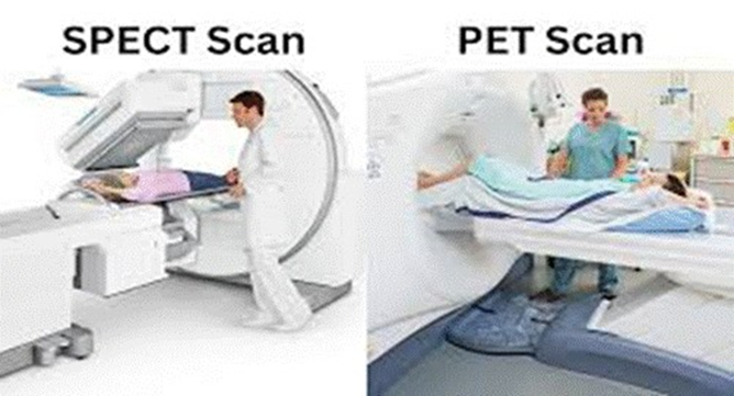
Figure 1: SPECT Scan and PET Scan
Imaging modality & Physics
|
Feature |
Technetium-99m (⁹⁹ᵐTc) – SPECT |
Fluorine-18 (¹⁸F) – PET |
|
Decay mode |
Gamma emission (140 keV) |
Positron emission (β⁺, 0.64 MeV) |
|
Detection |
Uses a collimator for detecting gamma photons |
Detects annihilation photons (511 keV) from positron decay |
|
Resolution |
Moderate (6–10 mm) |
Higher (~3–5 mm) |
|
Sensitivity |
Lower than PET |
Higher sensitivity |
|
Attenuation |
More susceptible to attenuation |
Less attenuation due to higher energy |
|
Sensitivity |
Lower than PET |
Higher sensitivity |
|
Attenuation |
More susceptible to attenuation |
Less attenuation due to higher energy |
Table 1. Comparison of Features between Technetium-⁹⁹ᵐTc-SPECT and Fluorine 18 ¹⁸F-PET
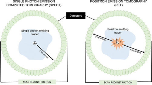
Figure 2. Scan Reconstruction of SPECT(L) and PET(R)
Radiopharmaceuticals & Applications
|
Feature |
⁹⁹mTc-SPECT Imaging |
¹⁸F-PET Imaging |
|
Common tracers |
⁹⁹mTc-MDP (bone imaging), ⁹⁹mTc-sestamibi (cardiac perfusion), ⁹⁹mTc-PSMA, ⁹⁹mTc-DMSA (renal imaging) |
¹⁸F-FDG (glucose metabolism), ¹⁸F-PS- MA, ¹⁸F-DOPA, ¹⁸F-NaF (bone imaging) |
|
Theranostic applications |
Used in SPECT imaging to select candidates for targeted radiotherapy (e.g., ⁹⁹mTc-PSMA for prostate cancer leading to ¹⁷⁷Lu-PSMA therapy) |
Used in PET imaging for staging and guiding targeted radiotherapies (e.g., ¹⁸F-FDG for metabolic tumors, ¹⁸F-PS- MA for prostate cancer) |
|
Organ/system focus |
Bone, kidney, heart, tumors |
Oncology (most cancers), neurology, cardiology |
Table 2. Comparison of Radiopharmaceuticals & Applications Features between Technetium-⁹⁹ᵐTc-SPECT and Fluo rine 18 ¹⁸F-PET
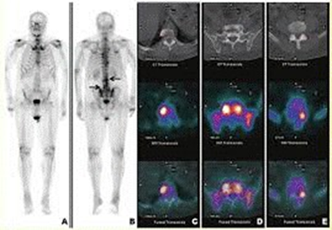
Figure 3. Evaluation of musculoskeletal sarcomas by using ⁹⁹ᵐTc-SPECT Imaging and ¹⁸F-PET Imaging [1] Radiation dose & Safety
|
Feature |
⁹⁹mTc-SPECT Imaging |
¹⁸F-PET Imaging |
|
Half-life |
6 hours (suitable for transport and use in nuclear medicine) |
110 minutes (shorter, requiring on-site or nearby cyclotron) |
|
Radiation dose |
Moderate |
Slightly higher due to higher energy photons |
|
Patient safety |
Safer for frequent scans |
Higher radiation exposure but still acceptable for clinical use |
|
Feature |
⁹⁹mTc-SPECT Imaging |
¹⁸F-PET Imaging |
|
Half-life |
6 hours (suitable for transport and use in nuclear medicine) |
110 minutes (shorter, requiring on-site or nearby cyclotron) |
Table 3. Comparison of Radiation dose & Safety Features between Technetium-⁹⁹ᵐTc-SPECT and Fluorine 18 ¹⁸F-PET 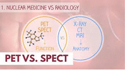
Figure 4. Nuclear medicine VS Radiology
Pros and Cons
|
Aspect |
⁹⁹ᵐTc-SPECT |
¹⁸F-PET |
|
Pros |
Widely available, cost-effective, good for bone and organ imaging |
Higher resolution, better sensitivity, superior quantification |
|
Cons |
Lower sensitivity, longer scan times, limited quantification |
Expensive, requires cyclotron, limited availability |
Table 4. Pros and Cons between Technetium-⁹⁹ᵐTc-SPECT and Fluorine 18 ¹⁸F-PET
Conclusion
• ⁹⁹ᵐTc-SPECT remains a cost-effective, widely available tool for molecular imaging, particularly in bone scanning and functional imaging.
• ¹⁸F-PET provides superior imaging quality, sensi tivity, and quantification, making it preferable for The choice between them depends on clinical needs, in frastructure, and cost considerations.
References
1. Garcia, R, Kim EE, Wong FC, and Korkmaz M, et al. “Comparison of fluorine-18-FDG PET and technetium-99m-MIBI SPECT in evaluation of musculoskeletal sarcomas.” J Nucl Med. 37(1996):1476-9.oncology, neurology applications. While SPECT is more accessible, PET is the gold standard for high-resolution, quantitative imaging.