Nabih Alkhouri, Mawia Karkoutly* and Nada Bshara
Department of Pediatric Dentistry, Faculty of Dentistry, Damascus University, Syrian Arab Republic
Received: 01 March 2024; Accepted: 08 March 2024; Published: 03 April 2024
Citation: Alkhouri Nabih, Karkoutly Mawia and Bshara Nada. “Effect of Sodium Hypochlorite on Marginal Microleakage of Composite Resin Restorations in Primary Teeth: An In Vitro Study” J Oral Dis Treat (2024): 102. DOI: 10.59462/JODT.1.1.102
Copyright: © 2024 Alkhouri et al. This is an open-access article distributed under the terms of the Creative Commons Attribution License, which permits unrestricted use, distribution, and reproduction in any medium, provided the original author and source are credited.
Background: This study aimed to evaluate the effect of 5.25% sodium hypochlorite (NaOCl) on marginal microleakage using class V composite restorations in primary teeth for different pretreatment times.
Methods: Class V cavities were prepared on the buccal surface of eighty primary canines. The cervical margin was located in the dentin, and the occlusal margin was in the enamel. Teeth were allocated into four groups (n=20): G1, Control, G2, pretreatment with NaOCl for 5 minutes, G3, pretreatment with NaOCl for 10 minutes, G4, pretreatment with NaOCl for 20 minutes. Rinsing or disinfecting of the cavities was performed before etching the cavities with 37% phosphoric acid. The total-etch adhesive system was used, then teeth were restored with nanohybrid composite. Specimens were immersed in 1% methylene blue solution for 4 hours following thermocycling, then bisected longitudinally in a buccolingual direction. Marginal microleakage was evaluated on the cervical and occlusal margins.
Results: The greatest marginal microleakage mean score (2.65 ± 0.75) was for group 4 on the cervical margins. There was a significant difference in marginal microleakage scores among the study groups on the cervical margins (p < 0.05). However, no statistically significant difference was detected on the occlusal margins (p = 0.25).
Conclusions: Longer exposure to NaOCl leads to marginal microleakage in cervical margins. The marginal microleakage was higher in the cervical margins.
Adhesion • Cavity disinfectant • Marginal microleakage • Primary teeth • Sodium hypochlorite
Marginal microleakage is a significant problem in composite restorations. It refers to the microscopic gaps between the composite material and the tooth structure, especially at the margins of the restoration. Gaps can occur due to several factors, such as polymerization shrinkage, incomplete cavity preparation, inadequate bonding, moisture contamination during placement, and the operator’s skill [1,2]. Marginal microleakage can lead to several sequelae, including recurrent caries, enamel demineralization, pulp irritation, and restoration failure. It can also compromise the aesthetics and longevity of the restoration. To minimize marginal microleakage in composite restorations, dentists adopt several techniques, such as proper cavity preparation, adequate bonding, incremental filling technique, isolation, and using flowable composite in the cervical area1. In addition, Dentists use advanced materials [3], such as low-shrinkage composites, universal adhesives, and bulk-fill composites [4,5], which are designed to reduce polymerization shrinkage and enhance bonding strength1. However, to date, the totaletch adhesive system is the gold standard in terms of bonding strengths [6].
Cavity disinfection before placing composite restorations is an important step to ensure the long-term success of the restoration. Disinfection aims to remove any remaining bacteria in the cavity, which can cause new decay and compromise the bond strength of the composite material. Therefore, Proper chemical disinfection adjunct to mechanical caries removal is essential to reduce the potential for marginal microleakage. There are several methods used to disinfect the cavity, including disinfectant solutions use, such as chlorhexidine, sodium hypochlorite (NaOCl), and hydrogen peroxide [7]. Sodium hypochlorite is an antibacterial and antiviral disinfectant commonly used in dental procedures. It’s also used as a bleaching agent due to its oxidizing properties. It’s known to be effective in eliminating microorganisms and reducing bacterial growth around restorations [8]. NaOCl can be used in pediatric dentistry in various ways as a root canal irrigant, pulpotomy agent, and disinfectant. The histopathological evaluation of NaOCl pulpotomy in primary teeth suggested that NaOCl decreases pulpal inflammation and necrosis. In addition, NaOCl induces dentinal bridge formation [9]. In addition, according to Bshara et al. [10], NaOCl solution yields satisfactory outcomes in terms of bovine pulp dissolution when compared to 2.2% NaOCl gel. Furthermore, primary teeth treatment with NaOCl does not affect the resin-dentin bonding strength [11]. NaOCl has been used as a root canal irrigant in pediatric endodontics for many years. It is a potent antimicrobial agent that can effectively dissolve necrotic tissue, disinfect the root canal system, and facilitate debris removal during chemomechanical preparation. It’s important to note that there is no specific time for NaOCl irrigation, and the clinician should adapt the timing to the individual child. Typically, the irrigation process can take from a few minutes to half an hour [12]. However, to date, the results of studies are controversial and scarce regarding the NaOCl effect on marginal microleakage in primary teeth whether for cavity disinfecting or root canal irrigation [13,14]. Thus, this study aimed to evaluate the effect of 5.25% NaOCl on marginal microleakage using class V composite restorations in primary teeth for different pretreatment times. The null hypothesis was that no statistically significant difference would be noted in the marginal microleakage between occlusal and cervical margins pretreated with NaOCl. In addition, no significant difference would be found in the marginal microleakage between different NaOCl pretreatment times.
Study design
This was an in vitro study. It was conducted at the Department of Pediatric Dentistry, Faculty of Dentistry, Damascus University, between December 2023 and January 2024. Ethical approval was obtained from the bioethics committee of Damascus University (N 1342/2023), and it was conducted in full accordance with CRIS Guidelines (Checklist for Reporting In-Vitro Studies). Written informed consent was obtained from patients before tooth donation.
Sample size calculation and study groups
The sample size was calculated using G* Power 3.1.9.4 software (Heinrich- Hein-Universitat-Dusseldorf, Germany; http://www.gpower.hhu.de/). Effect size f = 0.378/α err prob = 0.05/ Power (1-β err prob) = 0.80/ Number of groups = 4. Eighty sound primary canines were extracted for orthodontic treatment, cleaned out, and stored in a 1% chloramine solution (R1926000-500B, RICCA Chemical Company, Texas, United States). Primary canines were collected from patients undergoing serial extraction. Informed consent was obtained from all subjects’ legal guardian, and the study was conducted in full accordance with Helsinki Declaration 2013. Primary canines with physiologic root resorption of more than 1/3 root length, caries, enamel defects, fractures, or cracks were excluded. Teeth were randomly allocated into four groups:
Group 1: Control group, cavities were rinsed with distilled water (DW) (Pure Water, Pure Water Co., Aleppo, Syria) (n = 20).
Group 2: Cavities were pretreated with 5.25% NaOCl (CLOROX®, Oakland, California, United States) for 5 minutes (n = 20).
Group 3: Cavities were pretreated with 5.25% NaOCl for 10 minutes (n = 20).
Group 4: Cavities were pretreated with 5.25% NaOCl for 20 minutes (n = 20).
Specimens’ preparation
Class V non-beveled cavities were prepared on the buccal surface of the primary canines (depth, 1.5 mm; mesiodistal width, 3 mm; occlusogingival height, 2mm) by 330 carbide bur (Dentsply Maillefer, Tulsa, Oklahoma, United States). The cervical margin was located in the dentin (at the cementoenamel junction), and the occlusal margin was in the enamel. Rinsing or disinfecting of the cavities was performed before etching, then were etched with 37% phosphoric acid (N-Etch gel, Ivoclar Vivadent, New York, United States). NaOCl was applied using applicator brushes and stayed for 5, 10, or 20 minutes followed by drying for 5 seconds with an air syringe. Etching was done for 30 seconds in the enamel and 15 seconds in the dentin, then rinsed for 15 seconds, followed by drying for 5 seconds. The bonding agent (Teteric N-Bond, Ivoclar Vivadent, New York, United States) was applied and was cured for 20 seconds using LED dental curing light (Power Led, Foshan Jerry Medical Apparatus Co., Ltd, Guangdong, China) with an intensity of 1200 mW/cm2. Teeth were restored with nanohybrid composite (Tetric N-Ceram, Ivoclar Vivadent, New York, United States), then cured for 40 seconds, and diamond finishing bur was used (Dentsply Maillefer, Tulsa, Oklahoma, United States) for finishing the restoration. Primary canines were restored in DW at 37 °C for 24 hours. All specimens were subjected to a thermocycling aging process for 1500 cycles (5°C/55°C) with a dwell time of 60 seconds [15]. To ensure proper isolation, the apices of primary canines were sealed with sticky wax (Polywax, Bilkim Ltd. Co., Izmir, Turkey), and teeth surfaces were coated with two layers of nail varnish up to 1 mm from the cervical and occlusal margins of the cavities.
Microleakage testing
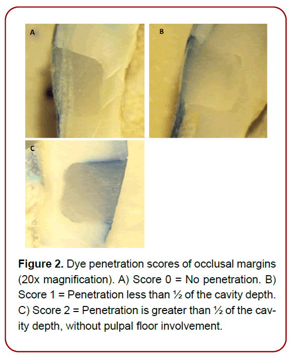
Specimens were immersed at room temperature in 1% methylene blue solution (Methylene Blue Saturated Aqueous Solution 1%, Science Lab Supplies, Texas, United States) for 4 hours, then washed and dried. Each primary canine was embedded in an acrylic resin block and bisected longitudinally in a buccolingual direction using a diamond disc bur (Dentsply Maillefer, Tulsa, Oklahoma, USA). Marginal microleakage was evaluated by two calibrated independent blinded outcome assessors on the cervical (Figure 1) and occlusal margins (Figure 2), using a stereomicroscope (Smart Optic, Seliga, Polska, Poland) at 20x magnification. Marginal microleakage was considered as the primary outcome measure. Dye penetration was evaluated using the following grading system (ISO/TS 11405) [15]:

Figure 1. Dye penetration scores of cervical margins (20x magnification). A) Score 0 = No penetration. B) Score 1 = Penetration less than ½ of the cavity depth. C) Score 2 = Penetration is greater than ½ of the cavity depth, without pulpal floor involvement. D) Score 3 = penetration with pulpal floor involvement.

Figure 2. Dye penetration scores of occlusal margins (20x magnification). A) Score 0 = No penetration. B) Score 1 = Penetration less than 1/2 of the cavity depth. C) Score 2 = Penetration is greater than 1/2 of the cavity depth, without pulpal floor involvement.
0 = No penetration.
1 = Penetration less than ½ of the cavity depth.
2= Penetration is greater than ½ of the cavity depth, without pulpal floor involvement.
3= Penetration with pulpal floor involvement.
Two operators performed the experiments and did the scoring. The Kappa coefficient of intra-examiner reliability was > 0.8. (Figure 1 and 2)
Statistical analysis
Statistical analysis was performed using IBM SPSS software version 22.0 (IBM Corp., Armonk, USA). Kruskal- Wallis test was applied for comparing among study groups, followed by the Mann-Whitney U test for pairwise comparisons at a significance level of p < 0.05.
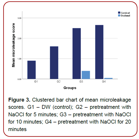
Marginal microleakage scores for occlusal and cervical margins are presented in Table 1. Groups 1 and 2 showed complete prevention of marginal microleakage on occlusal margins. The greatest marginal microleakage mean score (2.65 ± 0.75) was for group 4 on cervical margins (Table 2). Kruskal-Wallis test showed a significant difference in marginal microleakage scores among the study groups on the cervical margins (p < 0.05), but no statistically significant difference was detected on the occlusal margins (p = 0.25) (Table 2). A pairwise comparison between groups on the cervical margins is listed in Table 3. Mann-Whitney U test showed a significant difference between groups 1 and 3 and between groups 1 and 4 (p < 0.05). In addition, there was a significant difference between groups 2 and 3 and between groups 2 and 4. (Table 1-3) (Figure 3)

Figure 3. Clustered bar chart of mean microleakage scores. G1 – DW (control); G2 – pretreatment with NaOCl for 5 minutes; G3 – pretreatment with NaOCl for 10 minutes; G4 – pretreatment with NaOCl for 20 minutes
| Groups | n | Occlusal margins score | Cervical margins score | ||||||
|---|---|---|---|---|---|---|---|---|---|
| 0 | 1 | 2 | 3 | 0 | 1 | 2 | 3 | ||
| DW (control) | 20 | 20 | 0 | 0 | 0 | 8 | 9 | 0 | 3 |
| NaOCl (5 min) | 20 | 20 | 0 | 0 | 0 | 3 | 9 | 1 | 7 |
| NaOCl (10 min) | 20 | 16 | 0 | 4 | 0 | 1 | 1 | 5 | 13 |
| NaOCl (20 min) | 20 | 19 | 1 | 0 | 0 | 0 | 3 | 1 | 16 |
Table 1: Marginal microleakage scores for occlusal and cervical margins
| Cavity margin | Mean ± SD | df | p-Value | |||
|---|---|---|---|---|---|---|
| DW | NaOCl (5 min) | NaOCl (10 min) | NaOCl (20 min) | |||
| Occlusal | 0.00 ± 0.00 | 0.00 ± 0.00 | 0.40 ± 0.82 | 0.05 ± 0.22 | 3 | 0.25 |
| Cervical | 0.90 ± 1.02 | 1.60 ± 1.14 | 2.50 ± 0.83 | 2.65 ± 0.75 | 3 | < 0.001* |
Table 2: Comparison between groups for occlusal and cervical margins
| Groups | Mann-Whitney U | p-Value |
|---|---|---|
| DW vs NaOCl (5 min) | 129 | 0.056 |
| DW vs NaOCl (10 min) | 58 | < 0.001* |
| DW vs NaOCl (20 min) | 49.5 | < 0.001* |
| NaOCl (5 min) vs NaOCl (10 min) | 114 | 0.020* |
| NaOCl (5 min) vs NaOCl (20 min) | 101 | 0.007* |
| NaOCl (10 min) vs NaOCl (20 min) | 175 | 0.512 |
Table 3: Pairwise comparisons for cervical dye penetration
NaOCl is a well-known cavity disinfectant, but its efficacy in improving the marginal fit of composite restoration in primary teeth has not been exclusively studied [17].
Therefore, this study aimed to evaluate the effect of 5.25% NaOCl disinfectant on marginal microleakage in class V composite restorations in primary teeth for different pretreatment times.
Total-etch bonding systems involve etching the tooth surface with phosphoric acid before applying the bonding agent, which creates micropores that increase surface area for bonding and can result in higher bond strengths. Fifthgeneration bonding agents are also known as total-etch adhesives. In addition, they are known for their excellent bond strength, low technique sensitivity, and ease of use. Self-etch bonding, on the other hand, combines the etching and adhesives steps into one process. An acidic primer is applied to the tooth surface, which simultaneously etches and primes the surface for bonding. However, self-etch systems may have lower bond strengths and may not be suitable for some clinical situations. Fifth-generation bonding agents and self-etch adhesives have been shown to reduce microleakage compared to traditional techniques. However, self-etch adhesives have been shown to have slightly higher microleakage compared to fifth-generation bonding agents. This may be because self-etch adhesives do not create as rough a surface as total-etch adhesives, reducing the bond strength. However, it is critical to note that even with the use of fifth-generation bonding agents, microleakage can still occur [18,19]. In addition, they
may not be as effective in bonding in primary teeth as in permanent teeth due to the different composition structures [20]. Primary teeth have a thicker layer of enamel and a more porous dentin layer than permanent teeth, which makes it more challenging for the bonding agent to form a stronger bond [21]. Proper technique and placement of restorative materials can help to minimize microleakage and ensure long-lasting restorations [1]. The previous facts explain the use of a total-etch bonding agent.
The methylene blue dye penetration method was used due to its reliability and concise documentation [18,22]. Class V cavities preparations were performed as they are typically used to repair small cavities or defects. In addition, the configuration factor for class V restorations is usually low, as the cavity or defect is small and shallow [23].
In the current study, long pretreatment time was applied to match the time needed for root canal irrigation during endodontic treatment in primary teeth [12]. During endodontic treatment, NaOCl uses for irrigation could affect the bonding strength between the pulp chamber walls and composite restoration [24,25].
In the current study, no statistically significant difference in marginal microleakage scores among the study groups on the occlusal margins. However, there was a significant difference in marginal microleakage on the cervical margins.
This is because dentin is more vulnerable to microleakage due to its high permeability, which allows bacteria and fluids to penetrate easily. In addition, the enamel is a highly mineralized tissue due to its hydroxyapatite content [4,7,26]. Salama et al. [13] stated that the marginal microleakage in the cervical wall was superior to that in the incisal wall in primary teeth regardless of the disinfectant used. This result is in agreement with Bin Shuwaish et al. [7] study, which was performed on human premolars. In addition, NaOCl breaks up the long peptide chains in dentin and causes protein terminal group chlorination. This led to the fragment of the dentin organic matrix [27,28]. Zhang et al. [29] stated that the effect of NaOCl on collagen degradation is time-dependent. In addition, Zou et al. [30] suggested that the highest penetration for 6% NaOCl into the dentinal tubules (μm) was when applied for 20 minutes. This exceeded the penetration of acid etching leading to collagen network collapse, which is essential for bonding. Furthermore, Al Kurdi et al. [31] stated that primary dentin exposure to 5.25% NaOCl for 20 minutes negatively affects its microhardness. In addition, according to Moghaddas et al. [32], microleakage was higher in NaOCl-treated groups in comparison with non-NaOCltreated groups in human first permanent molars. Thus, the null hypothesis that no statistically significant difference would be noted in the marginal microleakage between occlusal and cervical margins pretreated with NaOCl, was rejected.
The main strength of the current in-vitro study is that the chemical and physical environment is tightly controlled. However, this study has limitations. First, sodium hypochlorite is unable to perform as accurately as in vivo exposure. Second, only sodium hypochlorite as a disinfectant agent was evaluated. Therefore, the results of the current study could be a prelude to conducting future clinical trials to match the oral cavity environment.
Based on the present study, longer exposure to NaOCl leads to marginal microleakage in cervical margins. Therefore, the marginal microleakage caused by NaOCl disinfectant is time-dependent. In addition, the marginal microleakage was higher in the cervical margins compared to the occlusal margins.
The authors have no conflicts of interest to declare.
N.A. collected data, extracted the data and performed the statistical analysis; M.K. wrote the manuscript; N.B. research concept and design, performed critical revision of the manuscript. All authors have read and approved the manuscript.
No grant or financial support was recieved from any governmental or private sector for this study.
Informed consent was obtained from all individuals, or their guardians included in this study.