Asaad Alsiddig Ahmed Mohammed2* and Lina Ahmed Hassaballa Ahmed1
1Department of Biology Faculty of Education University of Khartoum.
*Correspondence: Asaad Alsiddig Ahmed Mohammed, Department of Biology Faculty of Education University of Khartoum E-mail: asaadalsiddig@yahoo.com
Received: 15 Oct, 2025; Accepted: 31 Oct, 2025; Published: 14 Nov, 2025
Citation: Asaad Alsiddig Ahmed Mohammed and Lina Ahmed Hassaballa Ahmed. “Taxonomic Study Based on Wild Plant Species Related to Genus Datura L. in Sudan” J Environ Toxicol Res (2025):113 DOI:doi.org/10.59462/3068-3505.2.1.113
Copyright: © 2025 Asaad Alsiddig Ahmed Moham med. This is an open-access article distributed un der the terms of the Creative Commons Attribution License, which permits unrestricted use, distribution, and reproduction in any medium, provided the original author and source are credited
Abstract
This work is an attempt to make a comprehensive systematic study on the genus Datura at Shambat area, Khartoum State. It includes a brief description on climate, geology and topography of the study area.
Three species of Datura were studied, namely: Datura tatula, D. stramonium and D. innoxia. The systematic interrelationships between these species were determined using morphological, hitological, cytological and pollen grains characters.
Botanical names, synonyms and vernacular English and Arabic names have been presented. The study revealed that D. tatula is not a distinct species, but rather a variety of the species D. stramonium. The findings of this study have been illustrated using plates.
The cytology of this genus needs more elaborate and advance techniques. In this respect the author recommends the use of electron microscopy scanning to have a better view of chromosomes. It seems that there is a critical stage for identifying the various nuclear divisions and consequently chromosomal characterization.
Keywords
Plant Taxonomy, Datura innoxia, D. stramonium and D. tatula.
Introduction
The genus Datura is an important medicinal plant in all tropical areas. It has a variety of alkaloids that can be used for many medical and anaesthetic purposes. All the genus of Datura rich in tropane alkaloids, which have many medicinal and narcotic properties. Similarly, the genus is considered as a commercial source of hyos cine, a drug used for soothing hallucination. Moreover, its seeds have toxic alkaloids and even the least doses of these alkaloids are absolutely fatal. A number of tragic incidence have been reported in the Sudan as a re-sult of eating wheat or Dura flour containing Unknowingly the seeds of this genus. Besides its medici nal, anaesthetic and toxic effects, the genus Datura is also noxious weed of cultivation. The species of the genus are famous for hybridization, a phenomenon that adds a heavy task on taxonomists to determine the different varieties or hybrids resulting from these hybridizations.
The author of this work noticed obvious morphological in terrelationships between the species of the genus during the species collection, there was an apparent mixing of characters between the species of the genus. This obser vation encouraged the author to carry further work on this genus and the current study is an outcome of this obser vation.
This work is an attempt to classify the genus Datura on morphological, histological and cytological bases. The aims of this study are:
1. To identify the species of the genus Datura at Shambat area.
2. To determine interrelationships between the species and varieties of the genus. It is hoped that future studies may follow, complete and achieved what the author could not achieve in this study.
Study area
Site and location
The study area lies in the eastern part of Khartoum State, between latitude 15º 39´ N, and longitude 32º 31´ E on the eastern bank of the River Nile. It has an area of about 400km2.
Climate
Temperature
The coldest month is January with a mean temperature of 22.8Cº , whereas the hottest is June with a mean tempera ture of 33.4 Cº, during the short rainy season, the tempera ture falls to minimum of 30 Cº in August. The mean max imum air temperature ranges from 30.8 Cº in January to 42.2 Cº in May, while the mean minimum air temperature ranges from 14.2 Cº in January to 26 Cº in May. The mean daily range is large throughout the year. The lowest tem perature recorded in the area was 6 Cº in December in.
Rainfall
The rainy season extends through July, August and Sep tember. The normal mean annual total is 158 mm. There is considerable seasonal variation of rainy fall in this area both in a mount and distribution Rainfall could be below normal as in the year 1988. The duration of the conven tional rains varies from May to September with the peak in August. More than half of the annual total may fall in this month.
Wind
Wind blows from 2 directions, north and south. The wind speed varies from 8.p.h. (September to October) to 12 M.P.H. (February).
Relative humidity (R.H.)
The relative humidity is highest in the month of July to September with a peak in August, and lowest in the month of March to May. Regarding its geographical location, the study area has a tropical continental climate, which is usually hot and humid in summer, and mild and dry in winter, with a fairly market seasonal variation of temperature. The terminology (tropical continental) was first applied by [1]. When he classified the climate of the Sudan south of latitude 19º N. so this area lies in the central tropics where the year could be divided into 2 seasons: a wet summer and dry winter.
Geology and soil
The area is composed of ancient formation of cretaceous age, out cropping on the western bank of the Nile having a regional easterly dip of 0º - 5º with common high lo cal dips. Lithologically this formation, which is given the name of Nubian Series, is mostly sandstones, mudstones and ferruginous sandstones. The sandstones are made of coarse and fine particles of quartz. The ferruginous sand stones are composed of quartz grains and other accesso ry minerals in an ion oxide matrix, most probably limonite. Badly weathered feldspars, slender flakes of mica and cal cite crystal are common. The mudstones are impermeable compact formation formed of very fine grains. The base ment complex is made of recent deposits of Nile sands and silts. The sand is composed of angular grain of quartz mixed with particles of ion oxide and much biotite mica. The silts overlying sand are composed of very fine silt particles mixed with fine sand forming a light grey soil [1].
Topography
Most of the area is flat, interrupted by seasonal khors (ditches).
The Flora
This area is mostly under cultivation and hence the natural vegetation cover is continuously disturbed. Dense popu lations of plant growing naturally are only located in the idle land along the canals and in the very small areas that have not been brought under cultivation for many years. The flora of this area was studied by [2] who recorded 329 species of weeds. These weeds belonged to 60 families, the most dominant of which is Poacaea (Graminae), which is composed of 57 species. The most dominant species is Cynodon dactylon. Some of these weeds are poisonous, such as Datura spp. and Calotropis procera, and hence widely used in folk medicine.
In 1998, Bebawi studied the trees in the area and recorded 349 species belonging to 73 families. The most dominant family was Mimosaceae (29 species), and the most domi nant genus was Acacia (20 species). The indigenous Aca cia species on the list were Acacia seyal var. seyal, Acacia seyal var. fistula, Acacia mellifera and Acacia albida. The other Acacias were introduced and naturalized.
Material and Method
The plant materials were collected from Shambat area. This study is composed of 3 parts: morphology, anatomy and cytology each having its own method.
Methods used for the morphological study
The plant materials were collected through several trips to the study area, during the period of October 1998 to January 2001.
The collected plant specimens were air-dried, poisoned by mercuric chloride and mounted on herbarium paper for fu ture reference. Notes on habits, habitats, colour of flower, date of collection and distribution have been included. De scriptions of the family Solanaceae and the genus Datura are provided. Botanical names are updated. Synonyms, English names and vernacular Arabic names are given. A preliminary identification of the collected plant material has been done using [3]. This identification was later con firmed using [4-8].
Three species of the genus Datura were preliminary identi fied in Shambat area. These were namely: Datura innoxia, Datura stramonium and Datura tatula. A morphological de scription has been given for each species. The description covered vegetative, floral and fruit characters. (Figures 4, 6, 7, 8, 9)
Common uses have been given and these were mainly medicinal uses as the genus Datura represents an import ant medicinal plant. Reference has also been made to the most important alkaloid constituents of the genus. Plate’s illustrations have been used throughout the study.
Methods used for histological study
Preparation of plant materials
Cuts of about 2.5 cm. long were prepared using vegetative parts of roots, stems and leaves of the plant material. Flo ral and fruits parts were prepared using longitudinal and transverse sections of their respective parts.
Fixation
The prepared plant material was placed in vials (Table 1) containing Formaline Acetic Acid (FAA) which represented by the following formula:
| Ethyl alcohol (95%) | 50 cc | |
| Glacial acetic acid | 5 cc | |
| Formaldehyde (37 – 40%) | 10 cc | |
| Distilled water | 35 cc | |
| (Sass, 1958) | ||
Table 1. Concentrations of the vials solutions:
It was observed that the plant material cuts were fully im J Environ Toxicol Res 2025; Vol. 2(1) mersed in the fixative. The fixation was repeated 2 – 3 times until the solution become transparent. The plant was then labeled using a piece of paper and pencil.
Washing
The plant specimens were washed in distilled water for three times allowing a time of 20 minutes for each wash. The washing was very necessary inorder to a void the interference of acids in the staining process later on.
Dehydration
Series of different alcoholic concentrations (50%, 70%, 90% and 95%) were used for this purpose. In each con centration the plant specimens were left overnight or more to ensure complete dehydration.
Clearing
The plant specimens were cleared (Table 2) by using two mixtures as in the following table:
| Time | Mixture 1 Mixture 2 | |||
| Absolute alcohol Cedar wood oil Cedar wood oil Xylene | ||||
| 24 hrs | 100 | 0 | 100 | 0 |
| 3 hrs | 50 | 50 | 50 | 50 |
| 3 hrs | 25 | 75 | 25 | 75 |
| 24 hrs | 0 | 100 | 0 | 100 |
Table 2. Composition of clearing solutions:
Embedding in paraffin wax
A well-illustrated oven with thermostat-controlled electrical heating was used for infiltration. The oven was adjusted the day before embedding at 60 Cº. Three containers were put in the oven containing melted wax. The plant specimen was placed in closed vials containing 1:1 xylene and melted wax and put in the oven. The wax used in this step was W1 which is later replaced by a new pure wax called W2 after 45 minutes; W2 was replaced by a new pure wax called W3. The vials were left open so as to get rid of the xylene vapour.
Blocking
The equipment used in this process were: a heating source, a spatula, molds, strips of paper, a pencil and a trough cold water. The specimens were transferred from the vials to the mold containing pure melted wax. Each specimen was pressed gently against the peripheral part of the mold. The wax was left to consolidate.
Trimming
A scalpel was used for removing excess wax, and the re maining wax was left to support the plant specimen.
Sectioning
A rotary microtome, a brush, a razor blade, distilled water, trace, slides, a hot plate and cold atmosphere were used for this process. The stems sections were 14 microns thick while the leaves and the inflorescences were 9 and 12 microns thick, respectively. The ribbons were mounted on slides flooded with distilled water and placed on a hot plate so as to flatten the sections. The slides were removed from the plate and left to cool. The sections were then separated using a razor blade and each was mounted on a sep arate slide. The slides were returned to the hot plate and left to dry. They were then put in a tray and left overnight to ensure complete drying. The slides were then labeled using a diamond pen.
Staining
Two methods were adopted this respect:
a. Simple staining for the floral set as well as the fruit using hematoxylene mior.
b. Double staining for the vegetative parts roots, stems and leaves (Figures 11, 14, 15) using Safranine and Fast Green stains. Staining jars and glassware were used. The following solutions were also used: Xylene, different concentrations of ethyl alcohol (absolute alcohol, 95%, 90%, 70% and 50%), Safranine and Fast Green stains.
Procedures
The slides were put in staining jars and passed through the solutions. They were left for 4 minutes in Xylene and 2.5 minutes in absolute alcohol. They were then passed through alcoholic concentrations allowing a time of two minutes in each concentrations. The slides were then put in Safranine for 3 minutes and washed in the different alcoholic concentrations (50%, 70%, 90% and 95% absolute alcohol), respectively. Then the slides were immersed in Fast Green for 2 seconds, quickly washed in absolute al cohol and passed to Xylene. Cover slips and Canada bal sam were used to cover the slides. The slides were then taken to an oven at 60 Cº and left for 3 days.Procedures used in this section were after [9-11].
Methods used for the cytological study
Seeds of Datura were germinated in sand at different sea sons of the year (Figures 1, 2, 3). Root tips of the emerging radicles were collected and two methods were applied to examine their chromosomes. These methods were Aceto carmin Squash method and wax embedding.
a. Acetocarmin Squash method
The fixative was prepared immediately before collection. The collection was done directly in the field at different times of the day. The fixative used here was (1 part Glacial Acetic Acid: 3 part absolute Ethyl Alcohol).
Procedures: the root tips (Figure 14 & 15) were taken from the fixative and put into a heated 1 N HCL for 3 minutes in a test tube. Then they were washed in distilled water. A drop of basic Focsin was added and left for one minute. The root tips were then washed in distilled water. Each root tip was mounted. A drop of Acetocarmin was added and the slide was covered by a cover slip and pressed gently to squash the root tips. The squash was then examined under light microscope (X 100). The squash method is commonly used to determine the appropriate time of the collection and the cell stage where the chromosomes are quite visible [12].
b. Embedding in wax
The fixative used here was Formaline Acetic Alcohol (FAA) which was referred to the methods used for hitolog ical study and the same procedure used there was also followed here with minor modification. The root tips were placed in a petri dish containing the fixative and pressed horizontally between two slides to avoid any axis problems in the sectioning later on. In blocking, the root tips were placed horizontally at the bottom of the mold in the oven. Also, pre-section staining was conducted to distinguish between the root tips and the wax. The staining methods used in the histological study were also applied here to examine chromosomes. [9,10].
Results
Results of the morphological study
The genus Datura, which represents the scope of this study, belonged to the family Solanaceae. Here is a brief description of the family.
Family Solanaceae
Mostly herbs and shrubs. Leaves simple, exstipulate, al ternate. Inflorescence cymose or axillary solitary, flowers hypogenous, actinomorphic, hermaphrodite, petals 5, unit ed, valvate or contorted, stamens epipetalous, alternate, with the petals, ovary mostly 2 locular, becoming 4 locular in Datura, ovules numerous with axile placentation. Fruit capsule or berry.
Genus Datura L
Undershrubs or annual herbs. Leaves ovate, up to 9 in. long. Flowers axillary solitary, petals contorted, ovary 4 – locular.
There are apparently 3 species of Datura in Shambat. These are: Datura innoxia, Datura stramonium and Datura tatula.
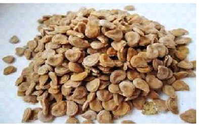
Figure 1: Seeds of Datura stramonium L.
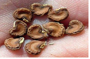
Figure 2: Seeds of Datura innoxia
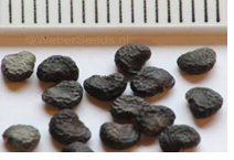
Figure 3: Seeds of Datura stramonium var.
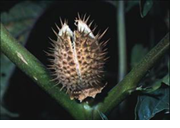
Figure 4: Mature fruit of Datura

Figure 5: Cross section in ovary of Datura spp
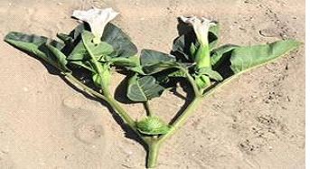
Figure 6: Datura innoxia Mill. whole plant
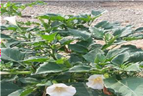
Figure 7: Datura stramonium whole plant
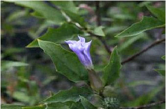
Figure 8: Datura stramonium var. tatula whole plant
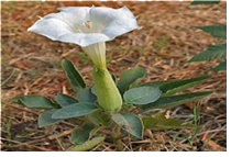
Figure 9: Sepals of Datura innoxia
Vernacular English and Arabic names
It is worth mentioning that the vernacular English name for the genus Datura is thorn apple whereas the vernacular Arabic name for it in Sudan is Sakaran.
Chemical constituents
All parts of the plant contain alkaloids. Hyoscine (C17 H21NO4) and hyoscine (C17H23NO3) are the common est besides some hyoscine and atropine (C17H23NO3). The plant material is more potent when than when dried.
Medicinal uses
Hyoscine has been employed to calm nervous irritation in hysteria and may be combined with purgative to avoid gripping. Atropine is used in the field of ophthalmology to dialate the pupil of the eye inorder to investigate certain diagnostic features of eye-sight disorder.
Toxicity
Poisonous material can be found in all parts of the plant, particularly the seeds. A tragic incidence occurred in the River Nile State when Datura seeds were harvested mis takenly with wheat grains. A bout thirty people lost their lives when unknowingly ate bread made from that wheat. It was noticed that herbivorous animals evade the plant because of its odour and harsh texture.
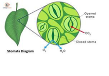
Figure 10: Stomata in Datura stramonium L
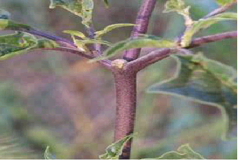
Figure 11: Stem of Datura innoxia
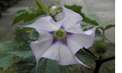
Figure 12: Flower of Datura innoxia
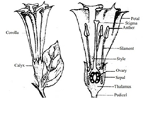
Figure 13: Anther & pollen grains in Datura innoxia
Figure 14: Roots of Datura innoxia
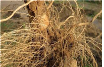
Figure 15: Root tips of Datura stramonium var. stramonium
Results of histological studies
This brief summary of anatomical features of the family Solanaceae:Trichomes in the family are diverse in forms, some being glandular while others aglandular. Leaves are usually dor siventral, but may be isobilateral in some species hairs, when present, have many kinds, spines, composed of elongated, lignifite cells, are present in certain species. Stomata may occur on both surfaces or often confined to the lower surface only (Figure 10). They may be ranucu laceous, cruciferous or caryophyllaceous. The mesophyll is sometimes mucilaginous. The wall of the palisade cells are sometimes thickened. Vascular bundles of the smallerveins are usually not accompanied by sclerenchyma. Crys tal occurs in various forms including solitary and clustered types. Collenchyma tissues are seen in the cortex layer of the stem and leaf midrib (Figure 11). A continuous xylem cylinder is observed in most genera. Intra-xylem is seen as a continuous cylinder frequently interrupted by patches of sclerenchyma. Pith is usually unlignified. Xylem vessels vary in size and xylem parenchyma is usually scantly. Xy lem fibers have bordered pits on the radial walls.
Hitological description of the genus Datura:
In addition to the above description given for the family, the genus Datura has these unique histological charac ters: stomata are generally cruciferous in shape; cells of the lower epidermis are set to contain chlorophyll. Crystal are prismatic in shape, the cluster crystals of Ca-oxalate mainly occur on the mesophyll while the solitary crystals occurnear the large vascular bundles. The larger vascular bun dles are bicollateral. stem cork is reported in the epidermal and sub-epidermal layer of the genus. Secondary phloem is usually devoid of fibers. Broad parenchyma rays may occur in mature secondary xylem. Interxylary phloem is recorded in the root of Datura stramonium. The genus Datura includes two major species D. stramonium and D. innoxia.
Tricellular uniseriate trichomes have been observed on the leaves, stems, petioles, peduncles and sepals of mature D. innoxia (Figure 9). However, no trichomes have been observed on the respective parts of D. stramonium which is found to be completely glabrous at maturity. The cells of the upper epidermis of the leaves of D. innoxia are narrower and elongated whereas those of D. stramonium are more less cylindrical. The palisade-spongy ration is 2:1 in D. innoxia compared to a ratio 1:1 in D. stramonium. The palisade of D. innoxia is darker in colour while that of D. stramonium is lighter in colour. The stomatal density on the lower surface of the leaves of D. stramonium is greater than that in D. innoxia. The subsidiary cell also differ in the two species. They are strongly wavy in outline in D. stramonium while they are less so in D. innoxia. The spongy cells of the mesophylls of the leaves of D. innoxia are compact while those of D. stramonium are more or less scattered and interrupted by numerous air-cavities. The spongy layer of the leaves of D. innoxia has more drusses than that J Environ Toxicol Res 2025; Vol. 2(1)of D. stramonium . Similarly the midrib parenchyma cells are more or less iso-diametric in D. innoxia while they are irregular in shape in D. stramonium. The epidermis of the stem of D. stramonium is two cells thick whereas that of D. innoxia is only one cell thick. The stem cortex collenchyma is 5 – 7 cells thick in D. stramonium while that of D. innoxia is 2 – 5 cells thick. Similarly the cortex parenchyma in D. stramonium is 7 – 10 cells thick whereas that of D. innoxia is 3 – 5 cells thick. The endodermal and pericyclic layers are conspicuous in D. innoxia while they are inconspicu ous in D. stramonium. The epidermal cells of the sepals of D. innoxia are elongated whereas those of D. stramonium are more or less cylindrical.The mesophyll layer of the sepals (Figure 9) of D. innoxia is 7 – 9 cells thick corresponding to 5 – 6 cells thick in D. stramonium.The pollen grains are carried on a long stalk from the wall of the another lobes in D. innoxia. In D. stramonium the pollen grains are carried directly on the wall of anther lobes (Figure 13).
Results of cytological study
The researcher has encountered a number of difficulties in this study respectively:Firstly, the seeds germination was very difficult and it took a lot of time to happened.Secondly, the stage where the division of chromosome is visible could not be determined in spite of the many at tempts that the researcher overtook to get this stage. It seems that there is specific time in the day when this division may occur and all trials to fix this time had failed. Thirdly, the chromosomes that have been detected in this study were not conspicuous enough to count or charac terize. However, some differentiation has taken place in Datura stramonium var. stramonium and anaphase and metaphase cell divisions could be identified.In Datura stramonium var. tatula the seeds failed to germi nate in spite of all effort to break seed dormancy through mechanical and chemical means.
As for Datura innoxia the seeds germinated but no differen tiation of chromosomes could be detected and the nuclear material appeared as one solid mass with any signal of nu clear division. Datura innoxia has 2n = 24 chromosomes, D. stramonium var. stramonium has 2n = 24 chromosomes while D. stramonium var. tatula has 2n = 25 chromosomes.
Discussion
In appears from the results of cytology that the genus Da tura is a very difficult genus to do any cytological study on it for the following reasons:
1 – Difficulty of seed germina tion, this because of water plug between the seed coat and the nucleolus and in the intercellular spaces of the spongy tissue within the hilum. [13].
2 – Difficult of determining the stage of cell division.
3 – Difficult of counting or characterizing chromosomes even if cell division occurred because of inconspicuous chromosome differentiation.
It seems that more advanced techniques are required in this respect. It could have been easier to do this study through electron microscopy but the only electron micro scope in the University of Khartoum has no functioning for several years till now 2025. It hoped that future studies will follow that utilizes more advanced techniques and elec tron microscopy to do a distinguished cytological study on this genus. However, a lot of information could be obtained from morphological and histological studies. These find ings have revealed a number of differences between the species of the genus Datura.
It is worth mentioning that the researcher could not get D. metel because it grows only in the Equatorial region. The remaining three species are characterized as follows: D. innoxia is morphologically quite different from the other species of Datura and hence it is considered as a distinct species.
There is striking morphological similarity between D. stra monium and D.tatula. The only difference is that could be noticed was the colour of stems, flowers and anthers. There was a great histological similarity between D. stra monium and D. tatula in all slides that had been examined. However, these species were found to be quite different from D. innoxia in many ways including presence of trichomes, shape of epidermal cells of the leaves, density of stomata and drusses in the leaves, the spongy palisade ratio, the shape and the thickness of the cortex parenchy ma cells, the thickness of the epidermal layer of the stem, shape and the mode of carriage of pollen grains in the anthers.
The anatomy of the root of all studied species of Datura revealed a striking similarity in structure and no significant differences were observed. Metacalf reported the pres ence of Interxylary phloem in the roots of D. stramonium. Hence, we can say:
1 – Datura innoxia is considered to be a distinct species.
2 - Datura tatula was found to be variety of Datura stra monium. There is a great morphological and hitological similarity between these two varieties. This finding is con firmed by [14] Palomino 1988 as mentioned earlier in this study.
Conclusions and Recommendation:
References